Niño con cachorro en brazos

Pathologies

Knowing more to be able to care better

The skin is the largest organ in the body and its main function is to act as the first line of defence against the outside world. For this reason, it is essential to keep it in a healthy state, which means identifying and treating any symptoms that may indicate an alteration in a timely manner. Knowing the most common dermatological pathologies in pets is essential to guarantee their well-being and ensure that they receive proper care.

PATHOLOGIES

Canine atopic dermatitis and feline atopic skin syndrome
Causes, Symptoms, Diagnosis, Treatment, and Prevention
Perro y gato

Introduction
Canine atopic dermatitis (CAD) and feline atopic skin syndrome (FASS) are chronic inflammatory skin diseases characterized by an overreaction of the immune system to environmental allergens. This triggers itching, irritation, and skin lesions.

Although the use of the term "atopic dermatitis" (AD) in cats is debated, this article refers to both conditions as AD.
AD is a highly prevalent condition that significantly impacts the quality of life of both species. This article addresses the causes, symptoms, and available treatments for managing this condition.

Causes
Although its etiology is multifactorial, the primary cause of AD is a genetic predisposition to react to certain environmental allergens. The most common allergens include:

  • Dust mites.
  • Plant and grass pollen.
  • Fungal spores.

In both species, the immune system reacts disproportionately when exposed to these allergens, causing skin inflammation. This inflammation is the primary cause of itching (pruritus), which leads animals to scratch, lick, or bite excessively, resulting in skin lesions.

Symptoms
Signs of AD in dogs and cats include:

  • Intense pruritus: Pets scratch, lick, or bite areas such as the abdomen, armpits, ears, and paws. In cats, this also includes the neck and back, often leading to skin wounds.
  • Redness (erythema): The affected skin becomes red, indicating inflammation and sensitivity.
  • Alopecia: Visible bald spots caused by constant scratching or licking, especially on the abdomen and paws.
  • Secondary infections: Wounds may become infected due to external factors and persistent licking.
  • Otitis externa: Inflammation of the ear canal with itching, redness, or discharge, common in dogs.
  • Specific lesions in cats: Miliary dermatitis (papules and crusts) and eosinophilic lesions such as ulcers, plaques, or granulomas in severe cases.
Some data
Image
Dermatitis atópica
15%
It is estimated that 10% to 15% of dogs worldwide suffer from atopic dermatitis.
Symptom
Pruritus (intense itching) is the most common symptom in dogs and cats.
Breeds
Certain breeds, such as the West Highland White Terrier and Labrador Retriever, are more prone to atopic dermatitis.

Diagnosis
The diagnosis of atopic dermatitis in dogs and cats includes:

  • Evaluating the clinical history (symptoms, age of onset, and seasonality).
  • Excluding other causes such as food allergies, bacterial or fungal infections, and ectoparasites.
  • Conducting intradermal or blood allergy tests to identify allergens.

Treatment
The treatment of atopic dermatitis aims to improve the animal's quality of life, alleviate pruritus, and prevent relapses. A multimodal approach includes:

  • Environmental control: Reducing allergens in the environment.
  • Systemic medications: Such as corticosteroids, immunomodulators, oclacitinib, and lokivetmab for dogs.
  • Personalized immunotherapy: For more severe cases.

The use of specific products with soothing and anti-inflammatory agents is essential to relieve symptoms, protect the skin, and enhance the animal’s well-being. Skinnia Calm offers an effective solution within this approach, providing immediate relief, strengthening the skin barrier, and maintaining adequate skin hydration.

Prevention

  • Environmental control: Regularly clean the environment to reduce allergens and use waterproof covers for beds and mattresses.
  • Skin care: Keep the skin hydrated with specific products and avoid excessive bathing with inappropriate products.
  • Proper nutrition: Provide a balanced diet rich in essential fatty acids to strengthen the skin barrier.
  • Veterinary check-ups: Regular monitoring to adjust treatments as needed.
Bibliography

Favrot, C., Steffan, J., Seewald, W., & Picco, F. (2010). A prospective study on the clinical features of chronic canine atopic dermatitis and its diagnosis. Veterinary Dermatology, 21(1), 23-31. 

Marsella, R. Atopic Dermatitis in Domestic Animals: What Our Current Understanding Is and How This Applies to Clinical Practice. Vet Sci. 2021 Jul 2;8(7):124. 

Halliwell, R. (2006). Revised nomenclature for veterinary allergy. Veterinary Immunology and Immunopathology, 114(3-4), 207-208

Scott, D. W., Miller, W. H., & Griffin, C. E. (2001). Muller and Kirk's Small Animal Dermatology. Elsevier, 6th ed.

Sousa, C. A., & Marsella, R. (2001). The ACVD task force on canine atopic dermatitis (X): guideline for treatment of canine atopic dermatitis. Veterinary Immunology and Immunopathology, 81(3-4), 285-296

Olivry, T., DeBoer, D. J., Favrot, C., Jackson, H. A., Mueller, R. S., Nuttall, T., ... & International Committee on Allergic Diseases of Animals. (2015). Treatment of canine atopic dermatitis: 2015 updated guidelines from the International Committee on Allergic Diseases of Animals (ICADA). BMC veterinary research, 11, 1-15.

Miller, J., Simpson, A., Bloom, P., Diesel, A., Friedeck, A., Paterson, T., Wisecup, M., & Yu, C. M. (2023). 2023 AAHA Management of Allergic Skin Diseases in Dogs and Cats Guidelines. Journal of the American Animal Hospital Association, 59(6), 255–284

Feline Dermatology. By Chiara Noli and Silvia Colombo (Eds). Springer Nature, Cham, Switzerland, 2020.

Related products
Skinnia Calm Gel Packaging
Gel

Bioadhesive transparent gel indicated to moisturise, protect and soothe the dry skin of small animals.

Seborrheic dermatitis
Causes, Symptoms, Diagnosis, Treatment, and Prevention
Perro Basset Hound en la playa

Introduction
Seborrheic dermatitis is a chronic skin condition affecting both dogs and cats, characterized by an imbalance in sebum production, which can result in dry or oily skin, flaking, and a foul odor. Although not always painful, this condition can cause significant discomfort for pets and lead to secondary infections if left untreated. This article explores the causes, symptoms, and available treatments for this condition.

Causes
Seborrheic dermatitis can be primary or secondary, and distinguishing between the two is key to proper treatment.

  • Primary seborrheic dermatitis: Genetic in origin, it is more common in certain dog breeds such as Cocker Spaniels, Basset Hounds, and West Highland White Terriers. Cats can also be affected, though it is less common.
  • Secondary seborrheic dermatitis: Can develop as a result of underlying conditions, including:
    • Allergies (environmental or food-related).
    • Bacterial or fungal infections.
    • Parasites such as fleas or mites.
    • Endocrine diseases like hypothyroidism in dogs.

Symptoms
Seborrheic dermatitis primarily affects the skin and coat and may present as either dry seborrhea (seborrhea sicca) or oily seborrhea (seborrhea oleosa). In some cases, pets may display a combination of both. Symptoms include:

  • Dry or oily skin: Depending on the type, the skin may be flaky and dry or oily and greasy to the touch.
  • Flaking (dandruff): Seborrhea often appears as small whitish flakes in the coat. In cats, this is commonly seen at the base of the tail.
  • Itching (pruritus): Causes excessive scratching and licking, increasing the risk of secondary infections.

Diagnosis
Diagnosing seborrheic dermatitis in pets involves a physical examination and symptom analysis. To determine whether it is primary or secondary, the veterinarian may perform tests such as:

  • Blood tests to rule out underlying diseases.
  • Skin cultures and microscopic evaluations to identify infections or abnormalities.
  • Skin scrapings to rule out mites.
  • Biopsies for deeper analysis in complex cases.
Some data
Image
Dermatitis seborréica icono
15%
Up to 15% of dogs may suffer from seborrheic dermatitis, particularly breeds like Cocker Spaniels and Basset Hounds.
Signs
Flaking (dandruff), unpleasant odor, and dry or oily coat are key signs.

Treatment
Treatment depends on whether the seborrheic dermatitis is primary or secondary, with a focus on addressing any underlying conditions in secondary cases.

  • Topical treatment: Key to alleviating symptoms, using products with keratolytic effects like Skinnia Repair, which exfoliates and softens the skin, removes dead cells, and maintains proper hydration to support regeneration.
  • Treatment for secondary infections: Topical antibiotics or antifungals may be required for infections.
  • Systemic treatment: May include omega-3 and omega-6 supplements to strengthen the skin and coat, and in severe cases, retinoids to normalize sebum production.

Prevention
While seborrheic dermatitis is not always preventable, certain measures can help reduce the severity of symptoms:

  • Regular bathing: Use specific shampoos to keep the skin healthy.
  • Frequent brushing: Removes excess dandruff and evenly distributes natural sebum.
  • Topical products: Maintaining hydrated and exfoliated skin with products like Skinnia Repair can improve skin condition and prevent relapses.
  • Allergy and underlying condition control: Managing these effectively can reduce predisposition to seborrhea, improving the pet’s quality of life.
Bibliography

Miller, W. H., Griffin, C. E., & Campbell, K. L. (2012). Muller and Kirk's Small Animal Dermatology (7th ed.). Elsevier Health Sciences.

Rosenkrantz, W. (2006). Practical applications of topical therapy for allergic, infectious, and seborrheic disorders. Clinical techniques in small animal practice, 21(3), 106-116. 

Borda, L. J., & Wikramanayake, T. C. (2015). Seborrheic dermatitis and dandruff: a comprehensive review. Journal of clinical and investigative dermatology, 3(2). 

Related products
Skinnia Repair Gel Packaging
Gel

Bioadhesive transparent gel with keratolytic effect indicated for smoothing and exfoliating the surface layer of the skin of pads and pressure points.

Hyperkeratosis
Causes, Symptoms, Diagnosis, Treatment, and Prevention
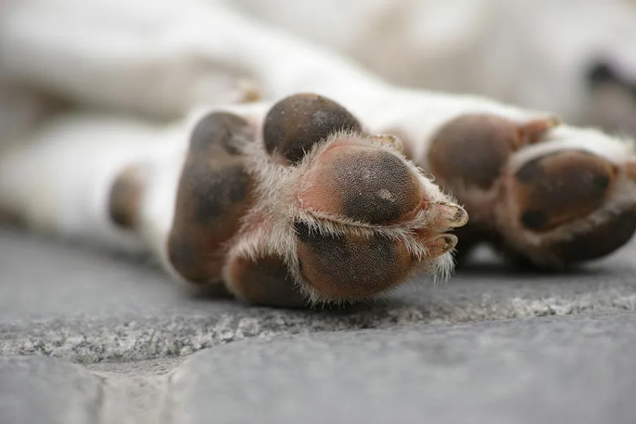
Almohadillas de un perro blanco

Introduction
Hyperkeratosis is a dermatological condition that mainly affects dogs and, less frequently, cats. It is characterized by abnormal thickening of the skin, particularly on the nose, pressure points, and paw pads. This condition can cause discomfort and even pain if not properly managed. This article explains what hyperkeratosis is, its causes, symptoms, and how to treat it to ensure your pet’s well-being.

Causes
Hyperkeratosis is a skin disorder caused by excessive production of keratin, a protein that forms the outer layer of the skin. This leads to thickening and hardening of the skin in specific areas, such as the paw pads (plantar hyperkeratosis) or the nose (nasal hyperkeratosis), making them dry, rough, and sometimes painful for pets.

Identifying the underlying cause is crucial for proper treatment. The most common causes include:

  • Genetics: Certain dog breeds, such as Labrador Retrievers, Bulldogs, and Cocker Spaniels, are more prone to developing hyperkeratosis due to hereditary factors.
  • Aging: As pets age, they may experience skin changes, such as increased keratin thickness.
  • Underlying diseases: Hyperkeratosis may be a symptom of other conditions, including:
    • Leishmaniasis in dogs.
    • Viral infections, such as canine distemper.
    • Autoimmune diseases, such as lupus.
  • Nutritional deficiencies: A lack of essential fatty acids or an inadequate diet may contribute to the development of hyperkeratosis.

Symptoms
The most common symptoms of hyperkeratosis include:

  • Thickening and hardening of the skin on the nose, paw pads, or pressure points.
  • Extreme dryness in affected areas.
  • Cracks that may cause pain.
  • Difficulty walking due to hardened paw pads.
  • In severe cases, bleeding or secondary infections.
Some data
Image
Hiperqueratosis icono
Where?
Hyperkeratosis primarily affects the paw pads and nose of dogs and cats.
What is?
Nasal hyperkeratosis can impair pets’ ability to perceive their surroundings as it compromises their sense of smell.

Diagnosis
A veterinarian diagnoses hyperkeratosis through a physical examination of the affected areas. If an underlying disease is suspected, additional tests such as bloodwork or skin biopsies may be performed to confirm the diagnosis.

Treatment
Hyperkeratosis can be effectively managed with appropriate treatment, improving your pet's quality of life and controlling symptoms.

  • Topical treatment: Essential for softening and removing excess keratin in the affected skin. Products like Skinnia Repair, a bioadhesive gel with salicylic acid and urea, help exfoliate and soften hardened skin, promoting regeneration. Hydrating products, such as specialized balms for paw pads and noses, are also helpful in preventing cracks and improving skin flexibility.
  • Treatment of underlying conditions: If hyperkeratosis is linked to an underlying disease, such as a viral or autoimmune condition, treating the primary disease is crucial. This may involve antiviral medications, antibiotics, or immunosuppressants.
  • Supplementation: Adding essential fatty acids to the diet can improve skin hydration and help regulate keratin production.

Prevention
To prevent and manage hyperkeratosis in pets, follow these care tips:

  • Regular application of hydrating and keratolytic products.
  • Protect paw pads on rough or hot surfaces and avoid walks during peak heat.
  • Maintain a balanced diet rich in essential fatty acids.
  • Consult a veterinarian if your pet shows signs of hyperkeratosis to ensure early diagnosis.
Bibliography

Robinson, K. (2015). Disorders of the paw. In BSAVA Manual of Canine Practice (pp. 327-338). BSAVA Library. 

Catarino, M., Combarros‐Garcia, D., Mimouni, P., Pressanti, C., & Cadiergues, M. C. (2018). Control of canine idiopathic nasal hyperkeratosis with a natural skin restorative balm: a randomized double‐blind placebo‐controlled study. Veterinary dermatology, 29(2), 134-e53.

Müller, G. H., Kirk, R. W., & Scott, D. W. (2012). Small Animal Dermatology (7th ed.). Saunders Elsevier.

Related products
Skinnia Repair Gel Packaging
Gel

Bioadhesive transparent gel with keratolytic effect indicated for smoothing and exfoliating the surface layer of the skin of pads and pressure points.

Flea Allergy Dermatitis
Causes, Symptoms, Diagnosis, Treatment, and Prevention
Cachorro que se rasca.

Introduction
Flea Allergy Dermatitis (FAD) is an allergic skin condition that primarily affects pets over one year old. It is commonly observed in dogs and cats with underlying skin diseases, such as atopic dermatitis. Pets with FAD often experience skin damage due to constant scratching, biting, or licking, which can lead to secondary infections.

Causes
FAD is caused by a hypersensitivity reaction to proteins in flea saliva. This condition is more common in warm and humid climates, which promote flea proliferation.

Often, flea control treatments are used inconsistently, leaving pets vulnerable to infestations during unprotected periods. To prevent this, it is important to apply treatments according to your veterinarian’s recommendations, ensuring year-round flea protection for your pet.

Symptoms

  • Intense pruritus (itching): Typically occurs in the caudal areas of the animal, especially near the tail.
  • Papules: Small bumps on the skin, often accompanied by excoriations from scratching.
  • Presence of fleas or flea dirt: Small dark specks visible on the pet’s coat.
  • Secondary infections: These may result from excessive scratching or licking and can include bacterial infections or yeast infections caused by Malassezia.
Some data
Image
Dermatitis alergica pulga icono
40%
It is estimated that FAD affects approximately 15-40% of dogs and 10-15% of cats in areas where fleas are prevalent.
30-50
Fleas lay between 30 and 50 eggs per day, meaning an infestation can rapidly grow in the pet’s environment if not controlled.
FAD
Flea Allergy Dermatitis (FAD) is one of the most common skin conditions in dogs and cats.

Diagnosis
The diagnosis of FAD is based on the pet’s clinical history, symptoms, and a detailed physical examination to identify signs of flea infestation, such as live fleas or flea dirt (small dark specks). In some cases, intradermal or serological tests may be conducted to identify allergic sensitivities, though these do not directly confirm the presence of fleas.

Treatment
Treating FAD starts with strict flea control, which involves treating all animals in the household and disinfecting the environment, as fleas can survive in carpets, bedding, and furniture.

To alleviate symptoms and improve the animal's well-being, topical treatments play a vital role. Products like Skinnia Calm help soothe irritated skin, reduce inflammation, and promote skin regeneration. In cases of severe itching, glucocorticoids or antihistamines may be necessary to control pruritus. Secondary infections should also be treated with antimicrobials as prescribed by a veterinarian.

Prevention

  • Regular flea control for all pets year-round.
  • Frequent cleaning of the environment to eliminate eggs and larvae.
  • Skin care: Use topical products like Skinnia Calm to strengthen the skin barrier.
  • Avoid infested areas and schedule regular veterinary check-ups.
Bibliography

Patel, A. & Forsythe, P. (2008). Saunders Solutions in Small Animal Practice. Veterinary Dermatology (1st edition). Saunders Elsevier.

Elsheikha, Hany & Wright, Ian. (2014). Flea infestations: epidemiology, treatment and control. The Veterinary Nurse. 5. 261-269.. 

Müller, G. H., Kirk, R. W., & Scott, D. W. (2012). Small Animal Dermatology (7th ed.). Saunders Elsevier.

Noli, C, Foster, A y Rosenkrantz, W, Veterinary Allergy, 2013; 1.ª edición, Wiley Blackwell, Nueva Jersey.

Related products
Skinnia Calm Gel Packaging
Gel

Bioadhesive transparent gel indicated to moisturise, protect and soothe the dry skin of small animals.

Contact Dermatitis
Causes, Symptoms, Diagnosis, Treatment, and Prevention
Perro Labrador Retriever en el bosque

Introduction
Contact dermatitis is an inflammatory skin reaction that occurs when the skin comes into contact with an irritant or allergen.
This condition can affect both dogs and cats and, while less common than other forms of dermatitis, it can cause significant discomfort and may lead to secondary infections if not properly treated.

Causes
Contact dermatitis in dogs and cats can be caused by:

  • Irritants such as detergents, disinfectants, or insecticides.
  • Plants, chemically treated grass, and synthetic materials found in collars, bedding, or toys.
  • Inappropriate topical products and metals like nickel, which may trigger allergic reactions.

Identifying and eliminating the irritant is crucial for preventing and managing this condition effectively.

Symptoms
The severity of lesions depends on the duration of exposure. Common symptoms include:

  • Erythematous areas: Skin with pink to red discoloration.
  • Alopecic areas: Hairless patches.
  • Pruritus (itching): Ranges from moderate to severe intensity.
  • Excoriations and crusts: Resulting from scratching.
  • Papules: Small raised bumps, and/or vesicles (small fluid-filled bumps) on the skin.
Some data
Image
Dermatitis por contacto icono
30%
Up to 30% of dermatitis cases in pets may be related to direct contact with irritants or allergens in their environment.
Abdomen
One of the most commonly affected areas in dogs and cats is the abdomen, as it is often more exposed and in contact with the ground and other surfaces.
Irritation
Contact dermatitis can appear within hours of exposure to an irritant, and symptoms can last days or even weeks if the causative agent is not removed.

Diagnosis
Diagnosis is based on the pet’s clinical history, observed symptoms, and any recent changes in habits or environment. It is critical to eliminate suspected irritants and monitor for symptom improvement.

To rule out other conditions, such as atopic dermatitis, parasites, skin infections, or food allergies, veterinarians may isolate the animal in a stainless-steel enclosure for 3-5 days. Improvement during this period confirms the diagnosis.

Treatment
Treatment begins with the identification and removal of the responsible allergen, which is fundamental for a good prognosis. If secondary infections caused by bacteria or Malassezia occur, specific antimicrobial treatments may be prescribed by the veterinarian.

To manage symptoms like pruritus and redness, soothing topical treatments are essential. Products like Skinnia Calm help reduce irritation, inflammation, and promote skin regeneration, significantly improving the pet’s well-being.

Prevention

  • Avoid exposure to irritants like detergents, disinfectants, or chemical products.
  • Use pet-specific products such as hypoallergenic shampoos and topical treatments.
  • Keep the skin hydrated and protected with treatments like Skinnia Calm.
  • Check the environment for plants or materials that could trigger reactions.
  • Monitor the use of accessories like collars or beds made from safe, non-allergenic materials.
Bibliography

Medleau, L., & Hnilica, K. A. (2017). Small animal dermatology: a color atlas and therapeutic guide. 4th ed. St. Louis, Mo., Saunders Elsevier.

Machicote Goth, G. (2011). Dermatología canina y felina (primera edición). Zaragoza: Servet

Ho, K. K., Campbell, K. L., & Lavergne, S. N. (2015). Contact dermatitis: a comparative and translational review of the literature. Veterinary dermatology, 26(5), 314-e67.

Related products
Skinnia Calm Gel Packaging
Gel

Bioadhesive transparent gel indicated to moisturise, protect and soothe the dry skin of small animals.

Food Allergies
Causes, Symptoms, Diagnosis, Treatment, and Prevention
Gato comiendo

Introduction
Food allergies are adverse immune reactions to certain foods or additives present in them. They affect both dogs and cats, although they are more common in dogs.
This condition can be challenging to identify and manage, as many symptoms can overlap with other skin or digestive diseases.

Causes
Food allergies in dogs and cats are primarily caused by specific proteins, both animal-based (e.g., beef, chicken, fish, or eggs) and plant-based (e.g., corn, wheat, or soy). Additives and preservatives in commercial pet foods may also trigger reactions in sensitive pets.

Prolonged exposure to the same food and genetic factors also play a role, with higher prevalence in breeds such as Labrador Retrievers, French Bulldogs, and Siamese cats. These allergies result from the immune system's response to certain ingredients, causing dermatological or digestive symptoms that require identifying and eliminating the allergen.

Symptoms
Food allergy symptoms are primarily dermatological and digestive.

Dermatological Symptoms

  • Non-seasonal pruritic dermatitis: Persistent itching throughout the year.
  • Skin lesions: Including hair loss (alopecia), excoriations, and crusts.
  • Secondary skin infections.
  • Erythematous-ceruminous otitis: Inflammation of the ears with redness and excess earwax. Approximately 25% of dogs with food allergies present this as their sole skin-related symptom.
  • In cats: Signs such as miliary dermatitis (small bumps and crusts on the skin) and lesions associated with the feline eosinophilic complex (ulcers and plaques related to allergies).

Digestive Symptoms

  • Vomiting.
  • Diarrhea.
  • Soft stools.
  • Increased frequency of defecation.
Some data
Image
Icono de Alergia alimentaria
30%
Food allergies account for 1-5% of dermatological conditions and 30% of allergic dermatitis cases in animals.

Diagnosis
Diagnosing food allergies involves:

  • Reviewing the clinical history to evaluate symptoms and patterns.
  • Excluding other potential causes, such as infections or parasites.
  • Using an elimination diet, considered the most effective method. This diet is maintained for at least 8 weeks to observe improvements, followed by controlled reintroduction of foods to identify the allergen.

Treatment
Treating food allergies in pets involves eliminating the causative allergen through an appropriate diet. To alleviate pruritus and irritation, topical products like Skinnia Calm are essential, providing effective local relief and improving skin condition.

In severe cases, veterinarians may prescribe glucocorticoids or antihistamines for temporary symptom control.

Prevention

  • Introduce varied diets early in life to avoid prolonged exposure to a single protein source.
  • Select high-quality foods, preferably hypoallergenic or with limited ingredients.
  • Avoid artificial additives or preservatives that may trigger sensitivities.
  • Consult a veterinarian before making significant dietary changes or introducing new protein sources.
  • Monitor for reactions after introducing new foods and act promptly if symptoms appear.
Bibliography

Wills, J., & Harvey, R. (1994). Diagnosis and management of food allergy and intolerance in dogs and cats. Australian Veterinary Journal, 71(10), 322–326

Miller, J., Simpson, A., Bloom, P., Diesel, A., Friedeck, A., Paterson, T., Wisecup, M., & Yu, C. M. (2023). 2023 AAHA Management of Allergic Skin Diseases in Dogs and Cats Guidelines. Journal of the American Animal Hospital Association, 59(6), 255–284. 

Medleau, L., & Hnilica, K. A. (2017). Small Animal Dermatology: A Color Atlas and Therapeutic Guide (4th ed.). St. Louis, Mo., Saunders Elsevier. 

Machicote Goth, G. (2011). Dermatología Canina y Felina (1st ed.). Zaragoza: Servet. 

Related products
Skinnia Calm Gel Packaging
Gel

Bioadhesive transparent gel indicated to moisturise, protect and soothe the dry skin of small animals.

Dermatophytosis (Ringworm)
Causes, Symptoms, Diagnosis, Treatment, and Prevention
Perro Border Collie

Introduction
Dermatophytosis refers to fungal infections of the skin, hair, or nails. The most common fungi responsible in dogs and cats belong to the genera Microsporum and Trichophyton. These fungi feed on keratin, a protein found in the outer layer of the skin, hair, and nails, allowing them to thrive and cause lesions.

Causes
Ringworm is highly contagious and easily transmitted through:

  • Direct contact with infected animals.
  • Indirect contact via contaminated objects such as brushes, bedding, and toys.
  • Exposure to contaminated surfaces, as the fungi can survive for long periods in the environment.

Certain factors increase the risk of dermatophytosis, including:

  • Hot and humid climates.
  • Immunosuppression in animals.
  • Young animals, as their immune systems are not fully developed.
  • Long hair, which provides an ideal environment for fungal growth.

Symptoms
The most common lesion is a circular hairless (alopecic) area, often red and crusty around the edges. While itching (pruritus) is mild to moderate, some pets may scratch the affected areas. Red, scaly, dry, and inflamed skin is also commonly observed.

In severe cases, the fungus can affect the nails, causing deformities and thickening, which complicates the infection.

Common sites for lesions include:

  • Limbs.
  • Face.
  • Ear flaps.
  • Tail.
Some data
Image
Icono de Tiña
15%
Approximately 10-15% of asymptomatic cats can carry the fungus and transmit the infection to other animals or humans without showing visible signs.
18 mos.
Fungal spores can survive in the environment for up to 18 months, requiring thorough disinfection of the pet’s surroundings to prevent reinfection.
Contagious
Dermatophytosis is highly contagious and can spread between animals and humans, making it a zoonotic disease that requires prompt attention.

Diagnosis
Diagnosing fungal skin infections involves the following tools:

  • Wood’s lamp examination: Detects the fluorescence of certain fungi, although a negative result does not rule out infection.
  • Microscopic examination: Identifies fungi in skin or hair samples.
  • Fungal cultures: Involves incubating samples in specific media to confirm fungal presence.

Treatment
Treatment for fungal skin infections includes both topical and systemic options:

  • Topical treatment: Essential for targeting localized infections using antifungal creams, lotions, gels, or shampoos. Products like Skinnia Calm can soothe irritation and support antifungal therapy by improving skin condition.
  • Systemic treatment: Recommended for severe cases or when topical therapy alone is insufficient. Oral antifungal medications such as griseofulvin, itraconazole, or ketoconazole should be administered under veterinary supervision.

Prevention

  • Hygiene: Regular baths with pet-specific products, avoiding excessive moisture on the skin and coat.
  • Avoid contact with infected animals: Limit exposure to pets or environments known to have fungal infections.
  • Disinfect the environment: Clean and disinfect pet bedding, blankets, brushes, and toys regularly.
  • Strengthen the skin barrier: Use topical products like Skinnia Calm to maintain healthy and hydrated skin, reducing the risk of infections.
  • Regular veterinary check-ups: Detect and treat early signs of skin irritation or infection.
Bibliography

Moriello, K. A., Coyner, K., Paterson, S., & Mignon, B. (2017). Diagnosis and treatment of dermatophytosis in dogs and cats: Clinical Consensus Guidelines of the World Association for Veterinary Dermatology. Veterinary Dermatology, 28(3), 266–e68. 

Machicote Goth, G. (2011). Dermatología Canina y Felina (1st ed.). Zaragoza: Servet. 

Related products
Skinnia Calm Gel Packaging
Gel

Bioadhesive transparent gel indicated to moisturise, protect and soothe the dry skin of small animals.

Pediculosis (Lice)
Causes, Symptoms, Diagnosis, Treatment, and Prevention
Perro Jack Russell rascándose

Introduction
Lice are external parasites that live on the fur and skin of animals. These small, wingless insects feed on either skin or blood, depending on the type of lice. The most common types of lice in pets are:

  • Chewing lice (Mallophaga): Feed on skin cells and hair, primarily affecting dogs.
  • Sucking lice (Anoplura): Feed on blood and are more common in cats.

It is important to note that lice are species-specific, meaning lice from pets do not infest humans and vice versa.

Causes
Lice infestations in dogs and cats typically occur through direct contact with infected animals, as lice do not survive long off their host. Infested environments, such as contaminated bedding, blankets, or grooming tools, can also be sources of transmission, especially in overcrowded or unhygienic conditions. Pets that do not receive regular care are at a higher risk of infestation.

Symptoms
Pediculosis presents with itching and skin irritation, prompting animals to scratch, rub, or bite themselves. Common signs include:

  • A rough or unhealthy coat appearance.
  • Severe infestations of sucking lice: Can cause anemia.
  • Severe infestations of chewing lice in dogs: Rarely lead to anemia.
  • Constant irritation and skin bites may result in restlessness, and wounds caused by lice feeding or the pet’s scratching may become infected.
Some data
Image
Icno de Pediculosis
2%
It is estimated that lice infestations affect around 1-2% of domestic pets in urban settings, with higher incidences in rural areas or in pets without regular preventive care.
Lice
Lice in dogs and cats are species-specific and cannot be transmitted between humans and animals or vice versa.

Diagnosis
Diagnosis is often made through direct observation of adult lice or their eggs on the animal, particularly in severe infestations. Microscopic examination of hair samples can also confirm the presence of eggs, aiding in diagnosis.

Treatment
Treatment for lice in pets involves the use of antiparasitic products available in various forms, including shampoos, spot-on pipettes, and collars. These are highly effective in eliminating and preventing infestations.

To relieve itching and irritation caused by lice, topical soothing products like Skinnia Calm are essential. These help to reduce inflammation, soothe discomfort, and support the regeneration of damaged skin.

Prevention
The best way to prevent lice infestations is through good hygiene and regular grooming checks. Key prevention measures include:

  • Using preventive products recommended by a veterinarian, such as antiparasitic collars or spot-on treatments.
  • Regularly washing the pet’s bedding, collars, and leashes, as well as cleaning areas where the pet spends time.
  • Maintaining clean and well-groomed fur to detect external parasites early.
Bibliography

Arther, R. G. (2009). Mites and lice: biology and control. Veterinary Clinics: Small Animal Practice, 39(6), 1159-1171. 

Little, S. E. (2021). Fleas and lice. In Greene's Infectious Diseases of the Dog and Cat (pp. 1324-1337). WB Saunders. 

Jackson, H. A., Marsella, R., & British Small Animal Veterinary Association. (2012). BSAVA Manual of Canine and Feline Dermatology (3rd ed.). British Small Animal Veterinary Association. 

Related products
Skinnia Calm Gel Packaging
Gel

Bioadhesive transparent gel indicated to moisturise, protect and soothe the dry skin of small animals.

Sarcoptic Mange (Scabies)
Causes, Symptoms, Diagnosis, Treatment, and Prevention
Cachorro de perro Labrador Retriever

Introduction
Sarcoptic mange, also known as “scabies,” is a highly contagious skin disease caused by the mite Sarcoptes scabiei var. canis. It primarily affects dogs but can also infect cats and, in some cases, humans. This infestation causes extreme discomfort in pets and can lead to serious health issues if left untreated.

Causes
Sarcoptic mange is caused by microscopic mites that burrow into the skin of the host to lay eggs and reproduce. This activity triggers intense itching and inflammation. The disease spreads easily through direct contact with infected animals or contaminated objects such as bedding and toys.

Symptoms
The hallmark symptom of sarcoptic mange in dogs is intense itching (pruritus). As the mites feed and damage the skin, the dog experiences severe itching and scratching. Other common symptoms include:

  • Hair loss (alopecia): Often around the ears, face, abdomen, and legs, resulting from scratching and biting.
  • Crusts and wounds: Caused by intense scratching and biting of the affected areas.
  • Red, inflamed skin (erythema): In affected areas.
  • Secondary infections: Due to constant scratching and skin damage.

In severe cases, the mange can spread over the entire body, and the condition may become chronic if not treated promptly.

Some data
Image
Icono de Sarna Sacóptica
15%
It is estimated that 3-5% of dogs in urban areas are affected by sarcoptic mange, with incidence increasing to 15% in rural areas or high-risk populations, such as shelters or overcrowded animal facilities.
2 wks
Symptoms may take up to six weeks to appear after exposure, allowing infected pets to spread the disease before showing visible signs.
Scabies
Sarcoptic mange is a highly contagious disease that primarily affects dogs but can also be transmitted to humans.

Diagnosis
Diagnosing sarcoptic mange involves:

  • Reviewing the clinical history.
  • Observing characteristic symptoms, such as hair loss, crusts, and redness on the ears, face, abdomen, and legs.
  • Performing skin scrapings, although mites may not always be detected.
  • Ruling out other causes of itching, such as infections or allergies.
  • Confirming the diagnosis by observing improvement after antiparasitic treatment.

Treatment
Treatment focuses on eliminating mites and relieving symptoms:

  • Antiparasitics: Medications such as ivermectin, milbemycin, or selamectin are effective in eradicating the mites.
  • Managing secondary infections: Antibiotics may be prescribed to treat bacterial infections resulting from scratching wounds.
  • Medicated baths: Help eliminate mites on the skin's surface and provide initial relief.
  • Topical products:
    • Skinnia Calm is essential for soothing irritated skin, maintaining hydration, and reducing discomfort, promoting rapid recovery.
    • Skinnia Wounds aids in wound healing and prevents infections caused by scratching.

In severe cases, antihistamines or corticosteroids may be prescribed to control inflammation and itching.

Prevention

  1. Avoid contact with infected animals.
  2. Regularly disinfect the environment: Clean and disinfect bedding, toys, and other items frequently, as mites can survive outside the host for weeks.
  3. Preventive antiparasitics: Consult a veterinarian about regular treatments to prevent mite infestations.
  4. Routine check-ups: Early detection and treatment can prevent the disease from spreading.
Bibliography

Müller, G. H., Kirk, R. W., & Scott, D. W. (2012). Small Animal Dermatology (7th ed.). Saunders Elsevier. 

Patel, A. & Forsythe, P. (2008). Saunders Solutions in Small Animal Practice: Veterinary Dermatology (1st edition). Saunders Elsevier. 

Bornstein, S., Mörner, T., & Samuel, W. M. (2001). Sarcoptes scabiei and sarcoptic mange. Parasitic diseases of wild mammals, 107-119. 

Curtis, C. F. (2012). Canine sarcoptic mange (sarcoptic acariasis, canine scabies). Companion animal, 17(8), 32-36. 

Related products
Skinnia Calm Gel Packaging
Gel

Bioadhesive transparent gel indicated to moisturise, protect and soothe the dry skin of small animals.

Skinnia Wounds Gel Packaging
Gel

Healing film forming gel indicated to protect and accelerate the natural healing process of the skin after surgery.

Ticks
Risk Factors, Symptoms, Diagnosis, Treatment, and Prevention
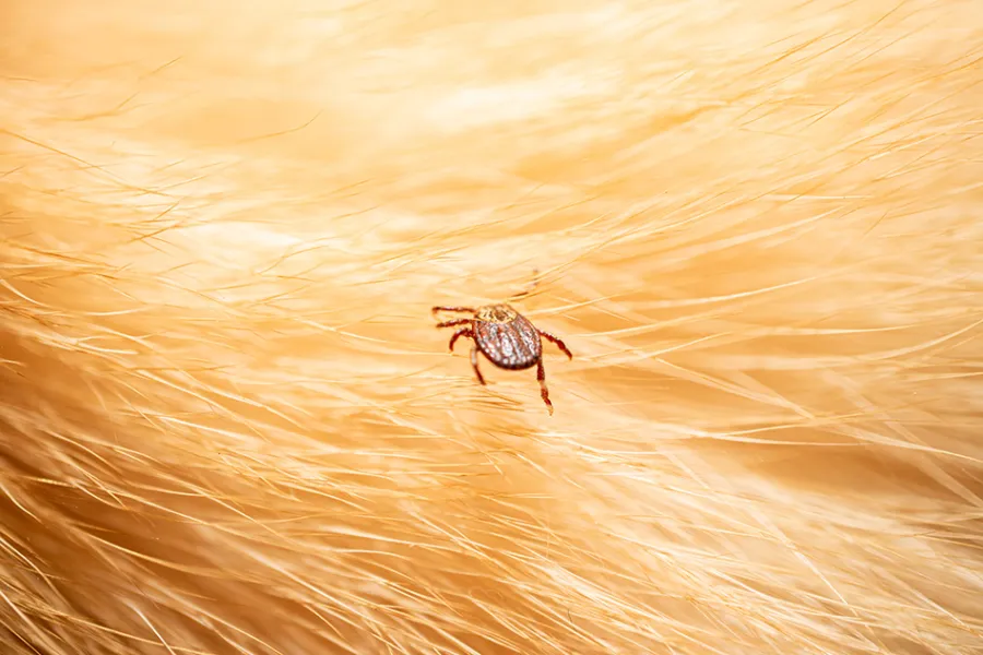
Garrapata en pelo de perro

Introduction
Ticks are arachnids that attach to the skin of animals to feed on their blood. Their presence on dogs and cats is more common in warm and humid weather. Besides being an annoyance to pets, ticks are vectors of various zoonotic diseases that can also impact human health.

Risk Factors
Ticks are commonly found in outdoor environments, especially wooded areas, tall grass, and shrubs. The following factors increase the risk of tick infestations:

  1. Outdoor exposure: Pets that spend a lot of time outdoors are more likely to encounter ticks.
  2. Warm, humid climate: Ticks thrive in warm and humid conditions, although in some regions, they can be active year-round.
  3. Lack of prevention: Pets without regular preventive treatments, such as spot-on applications, oral medications, or collars, are at higher risk.
  4. Long and dense coats: Ticks can hide more easily in thick fur, making detection more difficult.

Symptoms
Although ticks are small, their bites can lead to immediate and long-term symptoms, including:

  • Excessive scratching or licking: Ticks can irritate the skin, causing pets to scratch or lick more than usual.
  • Redness and swelling: Tick bites often result in inflamed, red areas, sometimes accompanied by rashes.
  • Lethargy or loss of appetite: In severe infestations, tick bites can lead to anemia and weakness in pets.
Some data
Image
Icono de Garrapata
2 years
Ticks can survive in the environment for up to two years without feeding, making them exceptionally resilient and challenging to eliminate.
3.000
A single tick can lay up to 3,000 eggs, meaning a small infestation can quickly escalate if not controlled.
15
Ticks can transmit more than 15 serious diseases, including Lyme disease, ehrlichiosis, and babesiosis, some of which can also affect humans.

Diagnosis
Diagnosing a tick infestation typically involves a physical examination, focusing on areas like the ears, neck, armpits, and between the toes. If symptoms like itching, redness, lethargy, or appetite loss are present, the veterinarian may suspect a tick-borne disease. In these cases, blood tests can detect infections caused by Borrelia, Ehrlichia, Anaplasma, or Babesia.

Treatment
Treating ticks involves both removal and symptom management:

  • Manual removal: Use tweezers to grasp the tick as close to the skin as possible and pull gently to ensure the entire tick is removed. Avoid squeezing the tick’s body to prevent infection.
  • Topical relief: Products like Skinnia Calm can soothe irritated skin, reduce inflammation, and promote the healing of tick bite sites.

Prevention
Preventing tick infestations is essential for safeguarding your pet’s health. Follow these steps:

  1. Use preventive products: Apply antiparasitic treatments, such as spot-on applications, oral medications, or collars, that protect against ticks for extended periods. Consult your veterinarian for the best option.
  2. Regular inspections: Check your pet’s coat frequently, especially after walks in wooded or grassy areas. Remove any ticks promptly.
  3. Avoid high-risk areas: During peak tick activity seasons, limit your pet’s access to areas with dense vegetation where ticks are common.
Bibliography

Medleau, L., & Hnilica, K. A. (2017). Small Animal Dermatology: A Color Atlas and Therapeutic Guide (4th ed.). St. Louis, Mo., Saunders Elsevier. 

Jackson, H. A., Marsella, R., & British Small Animal Veterinary Association. (2012). BSAVA Manual of Canine and Feline Dermatology (3rd ed.). British Small Animal Veterinary Association. 

Scott, D. W., Miller, W. H., & Griffin, C. E. (2001). Muller and Kirk's Small Animal Dermatology (6th ed.). Elsevier. 

Shaw, S. E., Day, M. J., Birtles, R. J., & Breitschwerdt, E. B. (2001). Tick-borne infectious diseases of dogs. Trends in parasitology, 17(2), 74-80. 

Estrada-Peña, A., Bouattour, A. J. L. C., Camicas, J. L., & Walker, A. R. (2004). Ticks of domestic animals in the Mediterranean region. University of Zaragoza, Spain, 131

Related products
Skinnia Calm Gel Packaging
Gel

Bioadhesive transparent gel indicated to moisturise, protect and soothe the dry skin of small animals.

Pyoderma
Causes, Symptoms, Diagnosis, Treatment, and Prevention
Perro salchicha lamiéndose

Introduction
Pyodermas are bacterial skin infections that can affect both dogs and cats, although they are more common in dogs. These infections can range in severity from surface-level infections to deep pyodermas involving multiple skin layers. Understanding the causes, symptoms, and treatment options for this condition is essential to keeping your pet's skin healthy and comfortable.

The term "pyoderma" refers to a bacterial skin infection, most often caused by Staphylococcus pseudintermedius, a bacterium normally present on the animal's skin that proliferates when the natural skin barrier is compromised.

Types of pyoderma in pets:

  1. Surface pyoderma: Affects the outermost skin layer, presenting with redness, itching, and small pustules.
  2. Superficial pyoderma: Involves the upper layers of the skin, including hair follicles. It commonly appears as pustules, redness, and hair loss in affected areas and is the most frequent form of pyoderma.
  3. Deep pyoderma: The most severe type, affecting deeper layers such as the dermis and subcutaneous tissue. It can cause abscesses, nodules, significant pain, and severe inflammation.

Causes
Pyodermas develop when the pet’s skin is weakened or compromised. Common causes include:

  • Allergies: Food, environmental, or contact allergies can irritate the skin, facilitating infections.
  • Parasites: Fleas, ticks, and mites irritate the skin and lead to scratching, increasing the risk of pyoderma.
  • Hormonal imbalances: Conditions like hypothyroidism or Cushing’s disease affect skin health and predispose pets to pyodermas.
  • Wounds or scratches: Any skin injury can become infected if not properly treated, particularly in sensitive or exposed areas.

Symptoms
The severity and extent of lesions depend on the type of pyoderma. Common signs include:

  • Papules: Small raised bumps on the skin.
  • Pustules: Raised bumps filled with pus.
  • Nodules: Larger skin elevations, deeper than papules.
  • Erosions and crusts.
  • Scaling: Flaky or scaly skin.
  • Abscesses: Localized pockets of pus.
  • Pigmentation changes in affected areas.

Other symptoms may relate to underlying causes, such as itching from allergies or lethargy from hormonal imbalances.

Some data
Image
Icono de Pioderma
50%
Up to 50% of skin infections in dogs are associated with pyodermas.
Pyoderma
Pyodermas are usually secondary to other health issues, such as allergies, hormonal diseases, or parasitic infestations.

Diagnosis
Diagnosing pyoderma begins with a thorough physical examination and review of the pet’s medical history. To confirm a bacterial infection and determine its depth, veterinarians may perform:

  • Cytology: Examining skin samples under a microscope to identify bacteria.
  • Bacterial cultures and sensitivity tests: Identifying the type of bacteria and determining effective antibiotics.
  • Additional tests: May include bloodwork or allergy testing to uncover underlying conditions.

Treatment
Treatment depends on the depth and extent of the infection and should be supervised by a veterinarian.

  • Topical treatments: Recommended for surface and superficial pyodermas. These may include antiseptics and antibiotics like mupirocin or fusidic acid.
  • Systemic antibiotics: Necessary for generalized, deep, or recurrent pyodermas. Antibiotics should be selected based on culture and sensitivity results.
  • Medicated shampoos: Containing chlorhexidine or benzoyl peroxide to cleanse the skin, reduce bacterial load, and alleviate symptoms.

Prevention
Preventing pyoderma involves maintaining good hygiene and addressing skin issues promptly. Recommendations include:

  • Parasite prevention: Use antiparasitic treatments to avoid infestations that can irritate the skin.
  • Proper grooming: Regular baths with pet-safe shampoos and maintaining clean fur and skin.
  • Balanced diet: Ensure the pet receives essential nutrients for healthy skin.
  • Veterinary check-ups: Especially important for pets prone to allergies or other skin problems.
Bibliography

Morris, D.O., Loeffler, A., Davis, M.F., Guardabassi, L., & Weese, J.S. (2017). Recommendations for approaches to methicillin-resistant staphylococcal infections of small animals: diagnosis, therapeutic considerations, and preventative measures. Vet Dermatol, 28: 304-e69. 

Machicote Goth, G. (2011). Dermatología Canina y Felina (1st ed.). Zaragoza: Servet. 

Santoro, D. (2023). Topical therapy for canine pyoderma: what is new?. Journal of the American Veterinary Medical Association, 261(S1), S140-S148. 

Bajwa, J. (2016). Canine superficial pyoderma and therapeutic considerations. The Canadian veterinary journal, 57(2), 204. 

Guaguere, E. (1996). Topical treatment of canine and feline pyoderma. Veterinary Dermatology, 7(3), 145-151. 

Malassezia Dermatitis
Causes, Symptoms, Diagnosis, Treatment, and Prevention
Perro West Highland White Terrier

Introduction
Malassezia dermatitis is one of the most common causes of skin and ear problems in dogs and cats. These infections are typically caused by Malassezia pachydermatis, a yeast that is part of the normal skin and mucosal flora in dogs. However, under certain conditions, this yeast can overgrow, leading to irritation and inflammation.

Causes
Malassezia is a fungus that naturally resides on the skin of dogs and cats. Under normal conditions, it does not cause harm, but certain factors can trigger overgrowth, leading to infection. Common triggers include:

  • Humidity and moisture.
  • Excessive oil production in the skin.
  • Weakened immune system.
    Some breeds, such as West Highland White Terriers, Dachshunds, Cockers, and German Shepherds, are more prone to these infections.

Symptoms
Malassezia dermatitis may be localized or generalized. Commonly affected areas include the face (ear canal and perioral skin), armpits, ventral neck, and distal limbs (interdigital spaces and nails). Symptoms vary but often include:

  • Intense itching: Pets may scratch constantly, especially in affected areas.
  • Redness and inflammation: Skin irritation and redness are common, especially on the paws, neck, and armpits.
  • Strong odor: Infected areas often emit an unpleasant odor, which is particularly noticeable in the ears.
  • Scaly or greasy skin: The skin may feel oily or show visible flakes.
  • Recurrent ear infections: Ear infections caused by Malassezia often involve dark discharge and a strong smell.
Some data
Image
Icono de Malassezia
20%
Around 15-20% of dogs with allergies also develop secondary infections caused by Malassezia due to constant scratching and weakened skin barriers.
Malassezia
Overgrowth of Malassezia produces a strong and distinctive odor, often described as similar to "toasted corn" or "rancid cheese."
Breeds
Breeds like West Highland White Terriers, Dachshunds, Cockers, and German Shepherds are predisposed to these infections.

Diagnosis
Diagnosis is performed by a veterinarian and includes:

  1. Clinical history and visible skin lesions.
  2. Cytology: Skin samples are examined under a microscope, revealing yeast cells with a characteristic "shoeprint" shape. A high number of these organisms confirms Malassezia dermatitis.
  3. Additional tests: May include blood and urine analysis to rule out underlying conditions contributing to yeast overgrowth.

Veterinarians also review any current treatments, particularly immunosuppressive medications such as corticosteroids, which can predispose pets to infections.

Treatment
Treatment involves a combination of therapies:

  • Topical therapy:
    • Antifungal and antiseptic shampoos and lotions containing ketoconazole or chlorhexidine are essential.
    • Products like Skinnia Calm can soothe redness and irritation, promoting comfort and healing.
  • Systemic antifungals: In severe cases, oral antifungals like itraconazole may be prescribed.
  • Addressing underlying causes: Treating allergies, hormonal imbalances, or other contributing factors is crucial to prevent recurrence.

Prevention
To reduce the risk of Malassezia dermatitis:

  • Keep skin clean and dry: Especially in moisture-prone areas like the paws and armpits.
  • Use appropriate care products: Choose pet-specific products that maintain a healthy skin balance.
  • Inspect and clean ears regularly: Particularly in breeds with floppy ears.
Bibliography

Machicote Goth, G. (2011). Dermatología canina y felina (primera edición). Zaragoza: Servet

Medleau, L., & Hnilica, K. A. (2017). Small animal dermatology: a color atlas and therapeutic guide. 4th ed. St. Louis, Mo., Saunders Elsevier.

Bajwa, J. (2017). Canine Malassezia dermatitis. The Canadian Veterinary Journal, 58(10), 1119. 

Bond, R. (2021). Malassezia dermatitis. In Greene's Infectious Diseases of the Dog and Cat (pp. 978-986). WB Saunders. 

Guillot, J., & Bond, R. (2020). Malassezia yeasts in veterinary dermatology: an updated overview. Frontiers in cellular and infection microbiology, 10, 79. 

Related products
Skinnia Calm Gel Packaging
Gel

Bioadhesive transparent gel indicated to moisturise, protect and soothe the dry skin of small animals.

Alopecia
Causes, Symptoms, Diagnosis, Treatment, and Prevention
Gato gris de pelo corto

Introduction
Alopecia refers to partial or complete hair loss in one or more areas of an animal’s body. While it can be a symptom of various dermatological conditions, it is commonly associated with diseases such as atopic dermatitis, where intense itching leads to excessive scratching and subsequent hair loss, or endocrine disorders like hypothyroidism. This article discusses the causes, symptoms, and treatments for alopecia in dogs and cats, along with tips for maintaining a healthy coat.

Causes
The causes of alopecia in pets are diverse and may include:

  • Parasites: Fleas, mites, and other external parasites are among the most common causes. Persistent itching can lead to self-inflicted hair loss.
  • Infections: Fungal infections (e.g., ringworm) and bacterial infections can result in localized hair loss.
  • Allergies: Food, environmental, or contact allergies can irritate the skin, causing excessive scratching and licking that leads to alopecia.
  • Hormonal imbalances: Disorders like hypothyroidism or Cushing’s syndrome disrupt hair growth and can cause localized or generalized hair loss.
  • Stress: Stress and anxiety, especially in cats, can result in compulsive grooming and hair loss.
  • Genetics: Certain breeds are predisposed to alopecia, particularly in specific areas of the body.

Symptoms
In addition to visible hair loss, alopecia may present with other symptoms that help identify the underlying cause:

  • Red or inflamed skin in affected areas.
  • Constant scratching or licking.
  • Scales, crusts, or dandruff on exposed skin.
  • Unpleasant odor if secondary infections are present.
  • Behavioral changes, such as restlessness or compulsive grooming.
Some data
Image
Icono de Alopecia
25%
In cats, up to 25% of hair loss cases are linked to excessive grooming due to anxiety or stress.
20%
Fungal infections, such as ringworm, account for approximately 20% of hair loss in cats.

Diagnosis
Diagnosing alopecia begins with a physical examination and review of the pet’s medical history. Additional diagnostic tests may include:

  • Skin cultures to identify fungal or bacterial infections.
  • Skin scrapings to detect parasites.
  • Blood tests to assess hormone levels and allergies.
  • Skin biopsies for more complex cases.

Treatment
Treatment depends on the underlying cause of alopecia:

  • Topical therapies: Specialized shampoos, soothing lotions, and fatty acid supplements improve skin and coat health. Products like Skinnia Calm are ideal for alleviating redness and discomfort in affected areas.
  • Environmental enrichment and play routines: Address stress-related alopecia by reducing anxiety in pets.
  • Veterinary care: More serious cases may require antifungal, antibiotic, or hormonal treatments as prescribed by a veterinarian.

Prevention
To minimize the risk of alopecia, prioritize your pet’s overall hygiene and health:

  • Use topical products to hydrate and protect the skin.
  • Feed a balanced diet rich in omega-3 and omega-6 fatty acids to improve the skin barrier and promote hair regeneration.
  • Regularly inspect and clean your pet’s skin and coat to detect issues early.
Bibliography

Heinrich, N.A., Eisenschenk, M., Harvey, R.G., & Nuttall, T. (2019). Skin Diseases of the Dog and Cat (3rd ed.). CRC Press. 

Paradis, M. (2012). An approach to symmetrical alopecia in the dog. In BSAVA Manual of Canine and Feline Dermatology (pp. 91-102). BSAVA Library. 

Hill, P. (2005). Clinical approach to alopecia in dogs-will the hair grow back?. 

Boeta, A. M. R. (2020). Diagnosis of alopecia in dogs and cats. Grupo Asís Biomedia SL. 

Related products
Skinnia Calm Gel Packaging
Gel

Bioadhesive transparent gel indicated to moisturise, protect and soothe the dry skin of small animals.

Solar dermatitis
Causes, Symptoms, Diagnosis, Treatment, and Prevention
Perro en la playa

Introduction
Solar dermatitis is an inflammation of the skin caused by prolonged sun exposure without adequate protection. It primarily affects areas with little to no hair, such as the nose, ears, abdomen, and back, particularly in pets that spend time outdoors. Light-skinned dogs, such as Bulldogs and Dalmatians, and hairless cats, like the Sphynx, are especially susceptible to solar dermatitis.

Symptoms
The severity of solar dermatitis symptoms depends on the duration of sun exposure and the pet’s skin type. Common symptoms include:

  • Redness of the skin: The affected area appears red and inflamed, similar to human sunburn.
  • Crusting or ulcers: In severe cases, the skin may develop crusts or ulcers, causing significant discomfort.
  • Itching and discomfort: Pets may excessively scratch or lick the irritated areas, exacerbating the condition.
  • Hair loss (alopecia): Hair may fall out in exposed areas due to sun damage.
Some data
Image
Icono de Dermatitis Solar
90%
Using pet-specific sunscreen can reduce the risk of solar dermatitis by up to 90%.
Sun
Sunlight is most intense between 9:00 a.m. and 3:00 p.m., increasing the risk of prolonged skin exposure.
3 times
Dogs with light-colored skin and hairless cat breeds are up to three times more likely to develop solar dermatitis.

Diagnosis
Diagnosis typically involves a review of the pet’s sun exposure history and a physical examination of the affected areas. In some cases, additional tests may be conducted to rule out other skin conditions.

Treatment
Treatment focuses on alleviating symptoms and preventing further damage:

  • Anti-inflammatory medications: Prescribed by veterinarians to reduce inflammation and pain.
  • Topical soothing products: Hydrating products like Skinnia Calm are effective in calming irritated skin and promoting healing.
  • Sun protection: Essential to prevent recurrence, including the use of pet-safe sunscreens and minimizing sun exposure during peak radiation hours. In severe cases, protective clothing or ensuring access to shaded areas is recommended.

Prevention
Preventing solar dermatitis involves minimizing sun exposure and protecting vulnerable areas:

  • Avoid peak sun hours: Limit outdoor activities during intense sunlight (9:00 a.m. to 3:00 p.m.).
  • Use pet-specific sunscreen: Apply to sensitive areas, such as the nose, ears, and abdomen, before outdoor activities.
  • Provide shade and fresh water: Ensure pets have access to shaded areas and hydration while outdoors.
Bibliography

Scarff, D. (2017). Solar (actinic) dermatoses in the dog and cat. Companion Animal, 22(4), 188-196. 

Scott, D. W., Miller, W. H., & Griffin, C. E. (2001). Muller and Kirk's Small Animal Dermatology (6th ed.). Elsevier. 

Burrows, A. (2012). Actinic (solar) dermatoses. In BSAVA Manual of Canine and Feline Dermatology (pp. 235-242). BSAVA Library. 

Scarff, D. (2007). Solar dermatoses in companion animals Part 1: Canine solar dermatoses. UK Vet Companion Animal, 12(3), 47-53. 

Related products
Skinnia Calm Gel Packaging
Gel

Bioadhesive transparent gel indicated to moisturise, protect and soothe the dry skin of small animals.